The Sensorimotor Hands-Brain Connection
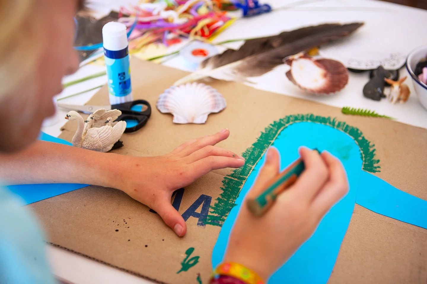
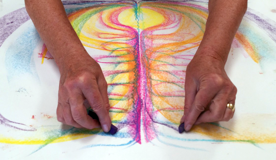
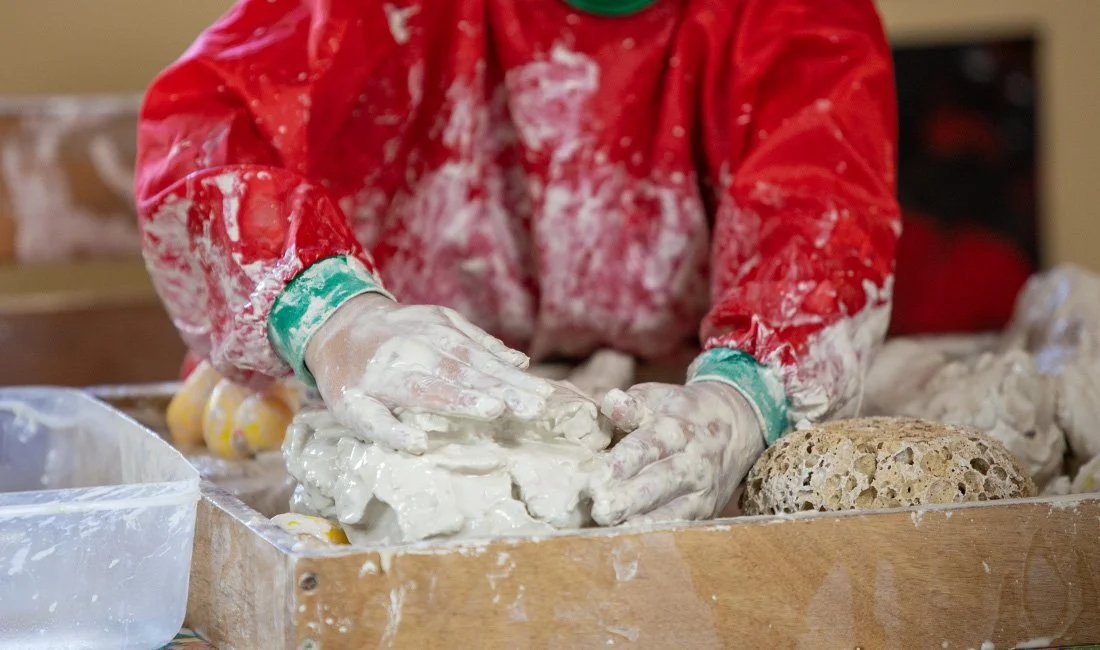
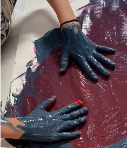
Haptic Perception describes touch-perception. While all artists and art therapy clients work with their hands, little attention has been given to the touch experience, when we work with crayons, paint, collage and clay, or play a musical instrument. However, in a society, where individuals spend increasing amounts of time online, children no longer play much outdoors, but instead press keyboards to engage with virtual realities, the need for psychological answers to this lack of touch connection becomes apparent. Not by accident body-focused approaches have been growing in importance in recent decade, and I am sure, so will haptic perception become an increasingly valuable therapeutic tool.
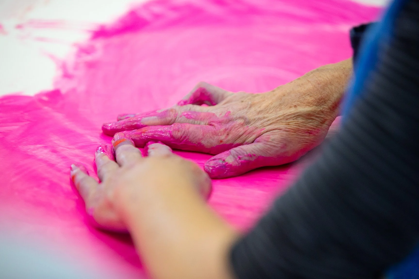
In his book called Touch, neuroscientist Linden[1] calls the skin a social organ through which we develop trust and bond with our caregivers. Our touch circuitry permeates every aspect of life. It literally makes us feel connected with family and friends. Linden even points out that doctors, who touch their patients get higher health care ratings and waiters higher tips, when they physically connect with their patrons. Children can develop normally without a sense of sight, sound, or smell. Yet without touch they grow up to be emotionally and socially dysfunctional; we could witness distressing evidence of this when images of the Romanian children raised in severely understaffed orphanages during the 1980s reached the Western public.
Touch is communicated to the brain via two mayor pathways: the peripheral nervous system and the fascia. The peripheral nervous system connects in the brain two mayor divisions, the motor and the sensory division. The sensory division, for example is in charge of our exteroceptive five senses perceiving the surrounding world, and and an interoceptive branch communicates our felt sense, how we sense the inside of our body as balance, internal sense and proprioception. The motor division has an involuntary division which regulates our heart beat, breath and the multitude of pulsing motor impulses our cells make without us ever thinking about it. And then there is the voluntary motor division, where I can decide to get up and get myself a cup of tea.
The fascial network, commonly called connective tissue, is the largest and medically most under-appreciated organ. It is a fascinating holistic system that holds our entire body together. All our bones, muscles, ligaments and organs ‘float’ in a myofascial web. Structural Integration expert Thomas Myers compares the fascia to a grapefruit. Within the skin of the grapefruit we find each fluid filled segment consists of a double walled membrane. Each slice is filled with many more tiny, softer, double walled membranes, each of these filled with juice.[2] In the human body “the fascial bags organize our ‘juice’ into discrete bundles, resisting the call of gravity to pool at the bottom.”[3] Our bones float inside this fascial web similar to the pips inside the grapefruit. What structurally holds us up is not the skeleton and the muscles and ligaments, but this intricate fascial net that encases everything in our body. We can easily prove this by trying to make a skeleton stand up. It won’t.
The fascia network reacts to touch through a variety of sensors that are located in a relatively shallow layer of the skin, and they answer to even tiny indentations produced by textured surfaces. “The fascial network is the largest sense organ in the body, dwarfing the eyes or ears in its rich diversity and proliferation of primarily stretch receptors. These sensory nerves frequently outnumber their motor compatriots in any given peripheral nerve by nearly 3:1.”[4]
Even light pressure onto in the fascial net is communicated across the entire system at lightning speed. Touch is essentially pressure. All injuries are stored in the fascia and leave imprints or even tear the membranes. The membranes respond to trauma and injuries by becoming stiffer and stiffer, which we then witness as bracing patterns such as a frozen shoulder and scars.
![Sensory and motor homunculus models at the Natural History Museum, London.[5]](https://images.squarespace-cdn.com/content/v1/5b162b70506fbe176d4215e0/1596013249696-O263O99D4FPQG1I2MJDW/Sensory+and+motor+model)
Sensory and motor homunculus models at the Natural History Museum, London.[5]
If we consider the ‘real estate’ the hands take up in the neocortex we find that the human hand and the mouth receive disproportionate representation in the brain. If we look at the homunculus models exhibited in the Natural History Museum in London depicting how the cortex relates to different parts of the body, it becomes apparent how significant sensory and motor impulses are as perception tools. Neurosurgeons Wilder Penfield and Theodore Rasmussen showed in their research that we use an enormous amount of our brainpower for motor dexterity, represented in the huge hands, and in our speech pictured in the giant lips and tongue. Smell and sight are of lesser importance and we don’t have much mental hardware devoted to the rest of our body.[6] Linden calls this phenomenon a touch map. That a “map of the body surface exists in the somatosensory cortex.”[7]
How would it be, if we imagine our clients connecting with the art materials with such huge hands. It illustrates so vividly, how important haptic perception is, and at the same time it is astounding, that we have researched this sensorimotor brain-hand connection so little.
The hand-brain connection particularly developed through skilled motor impulses. According to neurologist Wilson, the fact that we have developed a left and a right brain hemisphere is also due to both hands engaging in different tasks when they are no longer used for moving on all fours, when we were apes.[8] When I open a jar, for example, my left hand will hold the jar in place, while the right hand will apply a learnt motor impulse to twist the lid. In addition, the most effective way to communicate to others in my tribe how to open a jar, is by showing them the movements I make. In Neanderthal, hunting and survival discoveries would have been communicated through mimicry, hand-gestures, and the mirror neurons. A process that required the cortex to grow in order to store this new knowledge over a couple million years. Penfield’s homunculi shows with clarity, how strong the hand-brain connection is.[9] Without active touch experiences with our hands, the cortex will not become active in children.
Wilson describes how every child today will and needs to retrace these evolutionary motor skills in order to develop language. The baby’s signalling will become a signifying function. The verbal behaviour in the child undergoes a long metamorphosis during which words that were originally object attributes come increasingly to be manipulated and combined, just like real objects are manipulated and combined by the child. [10]
When a child piles building blocks on top of another pile, or groups beads in a cluster, or connects one place in in a painting with another through drawing a line, it exercises the cortical area that will later line up words like objects, group them into clusters and construct sentences. Hearing will only assure that the child of Chinese parents will not communicate in Italian, but the ability to form language skills depends on the child’s haptic motor skills. Child’s play is serious cognitive business.
My hope is that these insights will become increasingly integrated into the therapies we offer to our clients, in the context of learning difficulties and social connections. We can reach individuals through supporting haptic touch experiences with art materials, especially when their therapeutic needs are preverbal.
Bibliography
Linden, David J. 2015. Touch, the science of hand, heart and mind. Great Britain: Viking.
Myers, Thomas. 2014. November 20. https://www.youtube.com/watch?v=-uzQMn87Hg0.
Myers, Thomas. W. 2014. Anatomy trains; myofascial meridians for manual and movement therapists. Edinburgh, London, New York, Oxford, Philadelphia, St Louis, Sydney, Toronto: Churchill Livingstone Elsevier Ltd.
Penfield, Wilder. and Rasmussen, Theodore. 1950. renewed 1978. The Cerebral Cortex of Man: a clinical study of localization of function. University of Michigan. Macmillan.
Wilson, Frank. 1998. The hand; how its use shapes the brain, language and human culture. New York: Vintage Books.
© cornelia@sensorimotorarttherapy.com www.sensorimotorarttherapy.com
[1] Linden 2015, p. 7
[2] Myers 2014, p. 28
[3] Myers 2014, Youtube
[4] Myers 2014, p. 34
[5] Image from Shutterstock
[6] Penfield and Rasmussen 1950 renewed 1978
[7] Linden 2015, p. 62
[8] Wilson 1998, p. 147
[9] Wilson 1998, p. 15
[10] Wilson 1998, p. 193